44 diagram of the human eye without labels
Truth Giver of Humanity: What is the Human Race? - Blogger 06/05/2022 · These human variations will form in certain biomes or environments around the world and when humans migrated and conquered each other throughout human history; human variations, therefore, exist in more of a wide range of spectrums than as clear division. In fact, all variations exist in a spectrum from the most vestigial traits to the most functional trait that is … Blood vessels and nerves of the eye: Anatomy | Kenhub The human eye is a highly evolved structure of our anatomy and has many coexisting and interdependent elements. It is capable of moving and follow the objects along with accommodating to near and far; the eyes also can see in varying light, and in colour. Our two eyes working together give us stereoscopic vision, and depth perception.
Female Anatomy: Labeled Diagrams of the Reproductive System Reproductive anatomy aids with sexual pleasure, getting pregnant, and breastfeeding a baby. The urinary system helps rid the body of toxins through urination (peeing). The Female Reproductive System. Some people are born with internal or external structures that are ambiguous or characteristic of both male and female anatomy.

Diagram of the human eye without labels
Heart Diagram Labeled Igcse : The Human Eye Edexcel Igcse ... - Blogger This diagram shows the parts of the eye. …atrial septum may involve the atrioventricular valves and may be associated with incompetence of these valves. Sclera tapetum optic nerve blind spot lens vitreous humor iris cornea retina aqueous humor pupil. Your left is the heart's right, which is why the lefts and rights are swapped in the diagram. Free anatomy quiz worksheets: Learn anatomy faster! | Kenhub For beginners to the subject of human anatomy, the thought of having to learn hundreds of new structures can feel very overwhelming. Luckily, there are ways to make it easier. A great way to get familiar with the structures found within a particular region is to start by labeling human anatomy diagrams. Anatomical diagrams of the brain - e-Anatomy - IMAIOS Neuroanatomy : Brodmann areas, Broca area, Wernicke area. Three anatomical sections of the brain (axial, coronal and sagittal) close this chapter on the brain. Brain , Coronal section : Brain , Anatomy diagram. Numerous illustrations are available on the cerebellum, representation of cerebellar lobes, fissures, sulci and the vermis.
Diagram of the human eye without labels. Simple Diagram Of Human Eye With Labelling - Kaypa Khadzhiev Drag and drop the text labels onto the boxes next to the diagram. Click here to get an answer to your question ️ (a) draw a simple diagram of the human eye and label clearly the cornea, iris, pupil, ciliary muscles, . Eye diagram by firkin human eye diagram, diagram of the eye, eye parts,. Eye Diagram Quiz - ProProfs Quiz Try this amazing Eye Diagram Quiz quiz which has been attempted 5124 times by avid quiz takers. ... Label The Parts Of The Eye. People say that the eyes are the windows to a person's soul. ... How much did you get to understand about the human eye?... Questions: 8 | Attempts: 44861 | Last updated: Mar 22, 2022 . Sample Question. A is pointing ... Carburetor float diagram This video provides step-by-step instructions for replacing the float needle and seat on Briggs and Stratton small engines, commonly found in Toro, John Deer. Apr 13, 2022 · How a Carburetor Works – Float System The float circuit includes the float (brass, or nitrophyl), needle & seat, or float valve, bowl vent & float bowl. The float level and float drop should be measured to insure … Labeled imaging anatomy cases | Radiology Reference Article ... This article lists a series of labeled imaging anatomy cases by body region and modality. Brain CT head: non-contrast axial CT head: non-contrast coronal CT head: non-contrast sagittal CT head: angiogram axial CT head: angiogram coronal CT...
Understanding Different Eye Shapes: Which Do You Have? Hooded eyes: The lid appears smaller with this type of eye. This is because there is an extra layer of skin that is over the crease, which droops down. Protruding eyes: With this type of eye, the eyelids appear to project outward in the eye socket area. Upturned eyes: This type of eye has an almond shape. Data Visualization using Matplotlib - Towards Data Science 12/11/2018 · From the above diagram, the line that divides the box into 2 parts represents the median of the data. The end of the box shows the upper quartile(75%)and the start of the box represents the lower quartile(25%). Upper Quartile is also called 3rd quartile and similarly, Lower Quartile is also called as 1st quartile. The region between lower ... Anatomical Planes of Body - The Human Memory It is also known as Y-X plane or Frontal planes; the coronal plane divides the body into ventral (front) and dorsal (back) portions.This plane also gives a clear image of the posterior and anterior portions of the body. The coronal planes intersect the median plane at a 90-degree angle and show the anatomical body parts into front and back halves. Foot Anatomy and Common Foot Problems - Verywell Health Plus, the foot must be flexible to adapt to uneven surfaces and remain stable. Common foot problems include plantar fasciitis, bunions, flat feet, heel spurs, mallet toe, metatarsalgia, claw toe, and Morton's neuroma. This article provides an overview of foot anatomy and foot problems that come from overuse, injury, and normal wear and tear of ...
Diagram Maker | Online Diagramming and Design Solution Create eye-catching, informative diagrams without any design experience. Choose from a range of diagram templates to get started. Each diagram template is endlessly customizable, so you can make it as complex, concise or creative as you like. Venngage's free diagram maker lets you create engaging diagrams using unique icons and illustrations. Illustrations and diagrams of the 12 pairs of cranial nerves - IMAIOS This human anatomy module is about the cranial nerves. It consists of 15 vector anatomical drawings with 280 anatomical structures labeled. It is intended for the use of medical students working on human anatomy, student nurses, physiotherapists, electro-radiological technicians and residents - especially those working in neurology, neurosurgery, otolaryngology - and for any physician ... › heart › picture-of-the-heartHuman Heart (Anatomy): Diagram, Function, Chambers, Location ... Heart Tests. Electrocardiogram (ECG or EKG): A tracing of the heart’s electrical activity. Electrocardiograms can help diagnose many heart conditions. Echocardiogram: An ultrasound of the heart ... truthgiverofhumanity.blogspot.com › 2022 › 05Truth Giver of Humanity: What is the Human Race? - Blogger May 06, 2022 · In fact, the explanation given by racial supremacists for the differences between the brain size of certain human races is that humans were entered into environments that were not abundant in food and freshwater and adapted to these environments by growing larger brains that would help them come up with ways to survive without any physical ...

picture front of the eye without labels clipart - Clipground
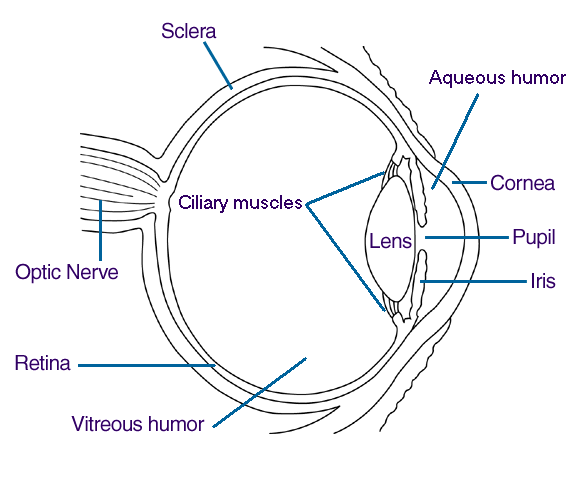
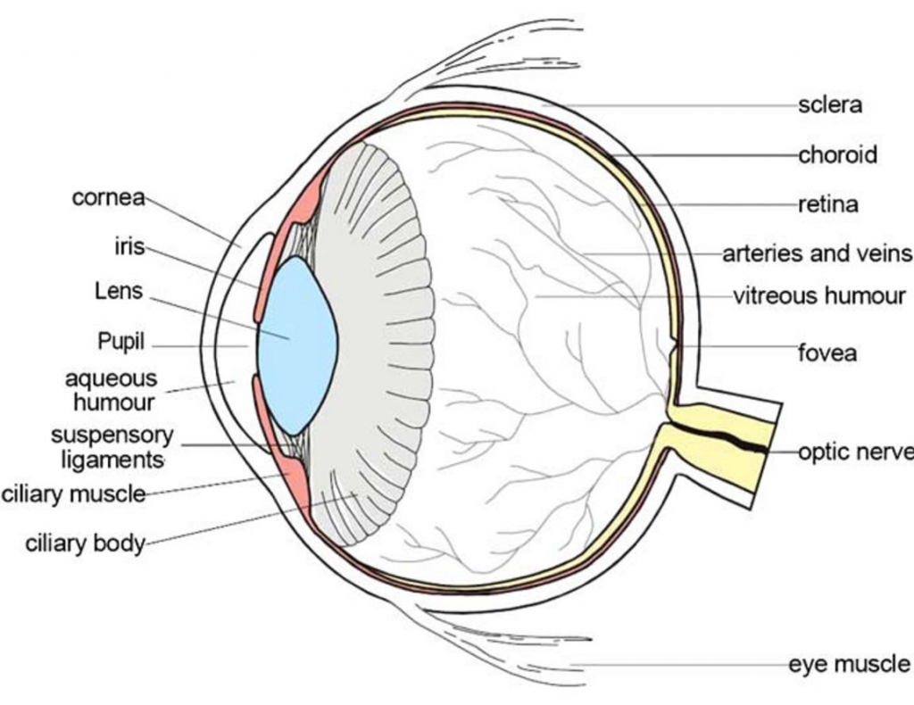
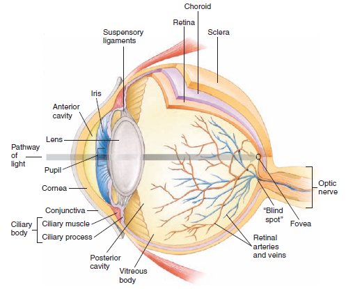
Anatomy of the eye: Quizzes and diagrams | Kenhub Take a look at the diagram of the eyeball above. Here you can see all of the main structures in this area. Spend some time reviewing the name and location of each one, then try to label the eye yourself - without peeking! - using the eye diagram (blank) below. Unlabeled diagram of the eye
Eye_Human
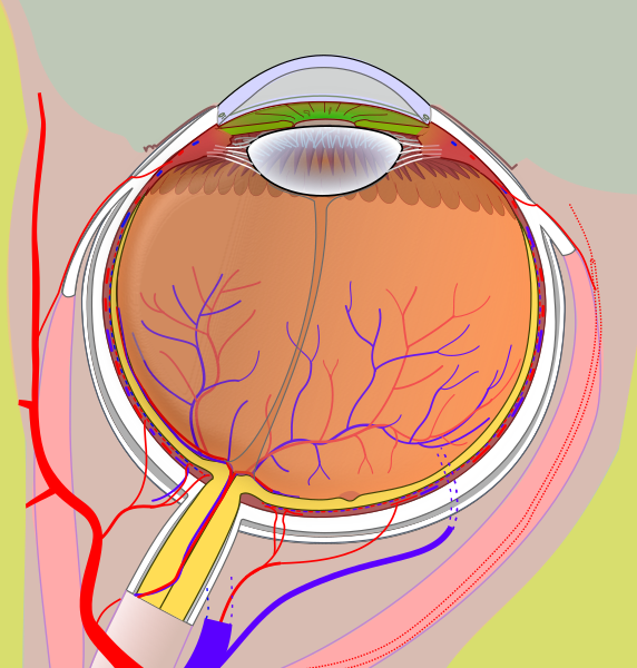
Human eye - Wikipedia The human eye is a sensory organ, ... Schematic diagram of the human eye. It shows a horizontal section through the right eye. The eye is made up of three coats, or layers, enclosing various anatomical structures. The outermost layer, known as the fibrous tunic, is composed of the cornea and sclera, which provide shape to the eye and support the deeper structures. The …
File:Diagram of human eye without labels.svg - Wikimedia Commons
rpxvu.plus-biznes.pl › carburetor-float-diagramCarburetor float diagram Mikuni Carburetor. category.Carburetors. Hwbnde HSR42 Easy Kit Carburetor 42mm, Works with Mikuni Carb Harley Davidson Evo Twin Cam 42-18 ️HSR42 42mm Carburetor ️Material: Total Zinc ️Fit for 1999-2006. this full -featured model meets all carb and epa regulations. horizontal throttle shaft through-bolt mounting design throttle bores with independent fuel circuits fixed high speed jet ...

File:Schematic diagram of the human eye en.svg - Wikipedia
Eye anatomy: Muscles, arteries, nerves and lacrimal gland - Kenhub The wall of the eyeball is three-layered; with the sclera as the outer layer (continuous with the cornea), choroid as the middle vascular layer (continuous with the ciliary body and iris ), and the retina as the innermost layer. You can study the anatomy of the eyeball in detail through this study unit. Anatomy of the eyeball Explore study unit

Module 1: Labeled Diagram of the Eye | Eye health | Pinterest | Activities
Parts of Stereo Microscope (Dissecting microscope) – labeled diagram ... The human brain is able to generate a 3-D image through the optical path and the angular offset. CMO or “Parallel style” stereo microscopes do not have double lenses; instead, the microscope has only one objective lens with a large diameter through which the light paths run for both the left and the right eye. This is why we called it ...
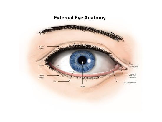
'External Anatomy of the Human Eye (With Labels)' Posters | AllPosters.com
Important Question for Class 10 Science Human Eye and Colourful World Draw labelled diagram of such lenses. (2020) Answer: (a) This condition is called presbyopia. (b) It happens due to gradual weakening of ciliary muscles and diminishing flexibility of eye lens due to agening. (c) It can be corrected by using bifocal lenses. Question 14. What eye defect is myopia?

38 best The Human Body images on Pinterest | Biology, Body diagram and ...
Consumer Updates | FDA - U.S. Food and Drug Administration 28/07/2022 · The site is secure. The https:// ensures that you are connecting to the official website and that any information you provide is encrypted and transmitted securely.

10 Best Images of Label Ear Diagram Worksheet - Blank Rock Cycle ...
Cells Diagram | Science Illustration Solutions - Edrawsoft Cells Diagram. Cells are the basic building blocks of all living things. The human body is composed of trillions of cells. Cells have many parts, each with a different function. Some of these parts, called organelles, are specialized structures that perform certain tasks within the cell. Drawing cells diagram helps you better understand your ...

17 Best images about Eye Anatomy on Pinterest | Eye anatomy, Human eye ...
Visible Human Project: anatomy atlas of male cadaver - IMAIOS The Visible Human Project is a fantastic tool that allows you to view almost all anatomical structures of the body. For didactic purposes and practice, we labeled one tenth of the possible structures to not overload the module. We deliberately set main names of bones, and summarily labeled the brain, almost all muscles of the body are listed.

picture front of the eye without labels clipart - Clipground
Anatomy of lower extremity - e-Anatomy - IMAIOS A diagram shows the various inguinal lymph nodes (lymphatic ganglia). The chapter on the innervation of the lower limb presents diagrams of the lumbosacral plexus and its main nerve branches for the lower limb (lateral cutaneous nerve of the thigh, femoral nerve, sciatic nerve and posterior cutaneous nerve of the thigh and obturator nerve).
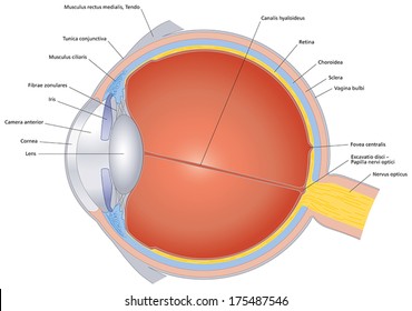
Diagram Of The Eye Not Labeled - Aflam-Neeeak
Blank ear diagrams and quizzes: The fastest way to learn - Kenhub It helps you to memorize the names and their locations, which in turn will aid you to remember their functions. Below, you can download both the blank ear diagram to make some notes, and then try labeling the ear using the unlabeled ear diagram. Good luck! DOWNLOAD PDF WORKSHEET (BLANK) DOWNLOAD PDF WORKSHEET (LABELED)
parts of the eyes clipart 20 free Cliparts | Download images on ...
Human penis - Wikipedia The human penis is an external male intromittent organ that additionally serves as the urinal duct.The main parts are the root (radix); the body (corpus); and the epithelium of the penis including the shaft skin and the foreskin (prepuce) covering the glans penis.The body of the penis is made up of three columns of tissue: two corpora cavernosa on the dorsal side and corpus …
Labelled Eye Diagram Human Eye Worksheet Answers - Diagram Media
The Lens: Anatomy, Function, and Treatment - Verywell Health The crystalline lens is a clear, biconvex layer of the eye that is made up mostly of proteins. As much as 60% of the lens mass is made up of proteins—a concentration higher than almost any other tissue in the body. 1 Four structures make up the crystalline lens: Capsule Epithelium Cortex Nucleus 2

Blank Ear Diagram | Human ear diagram, Ear anatomy, Ear diagram
en.wikipedia.org › wiki › Human_eyeHuman eye - Wikipedia The human eye is a sensory organ, ... Schematic diagram of the human eye. It shows a horizontal section through the right eye. ... Right eye without labels ...
picture front of the eye without labels clipart - Clipground
Anatomy of the Eye - Verywell Health View All. Cornea. Pupil. Iris. Crystalline Lens. Aqueous Humor. The human eye is an organ that detects light and sends signals along the optic nerve to the brain. Perhaps one of the most complex organs of the body, the eye is made up of several parts—and each individual part contributes to your ability to see.

Diagram Of Muscles In The Body / muscular system without labels ...
Anatomy and Structure of the Human Eye (With Diagrams) The iris is a flat, thin, ring-shaped structure sticking into the anterior chamber. This is the part that identifies a person's eye colour. The iris contains both circular muscles going around the pupil and radial muscles radiating toward the pupil. When the circular muscles contract, they make the pupil smaller.
Post a Comment for "44 diagram of the human eye without labels"