43 fluorescent labels and light microscopy
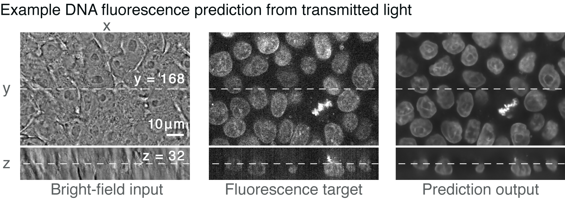
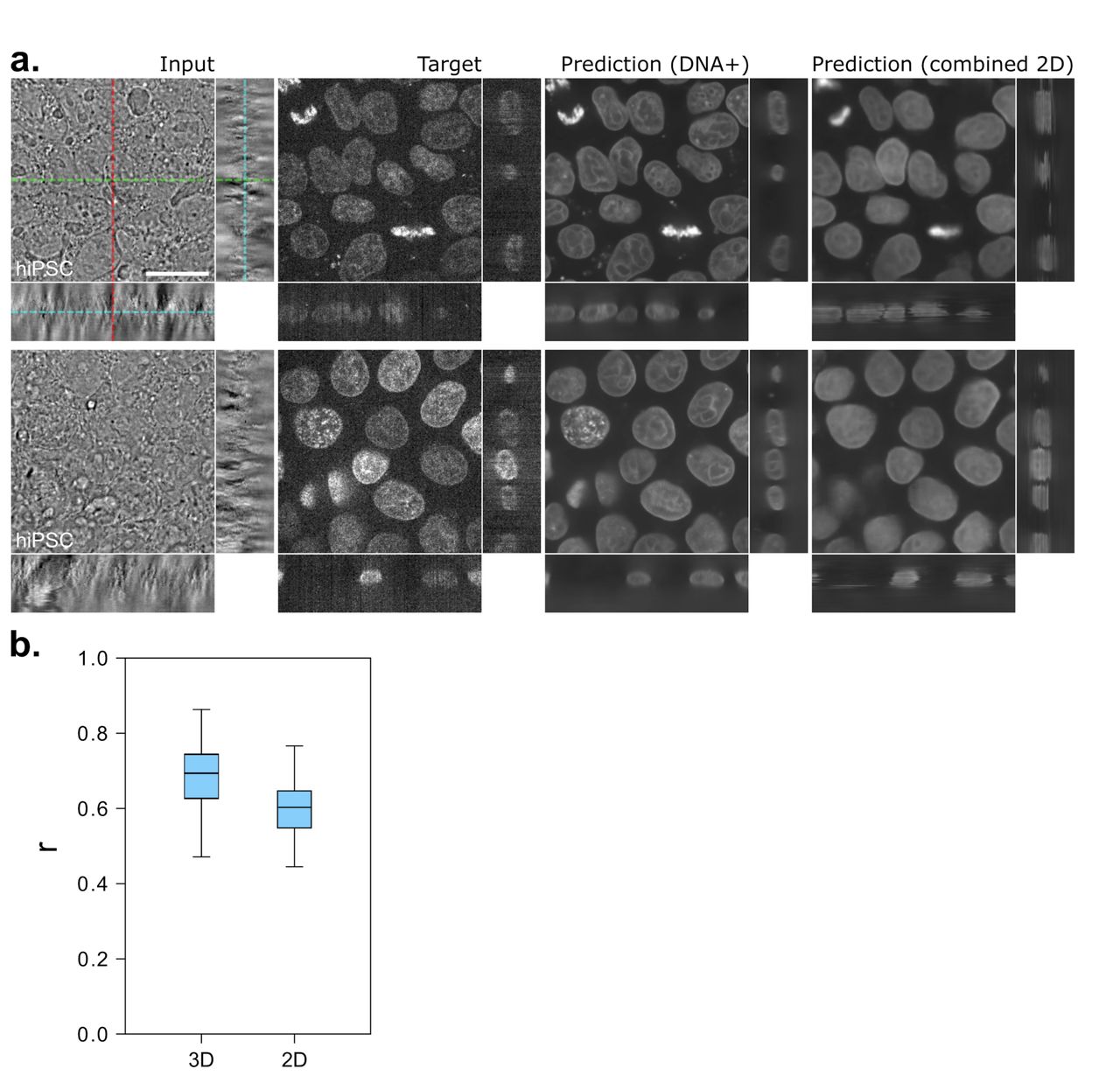
Different Ways to Add Fluorescent Labels - Thermo Fisher Scientific Fluorescent labeling methods can vary, As mentioned above, fluorescent labels usually comprise a fluorophore that is modified in a way that gives them specificity. In addition to linking fluorophores to various molecules, other types of modifications can give fluorophores novel characteristics. Label-free prediction of three-dimensional fluorescence images from ... We present a label-free method for predicting three-dimensional fluorescence directly from transmitted-light images and demonstrate that it can be used to generate multi-structure, integrated images. The method can also predict immunofluorescence (IF) from electron micrograph (EM) inputs, extending the potential applications. Publication types,
Light sheet fluorescence microscopy guided MALDI-imaging mass ... - Nature Light sheet fluorescence microscopy (LSFM) of optically cleared biological samples represents a powerful tool to analyze the 3-dimensional morphology of tissues and organs. ... In silico labeling ...
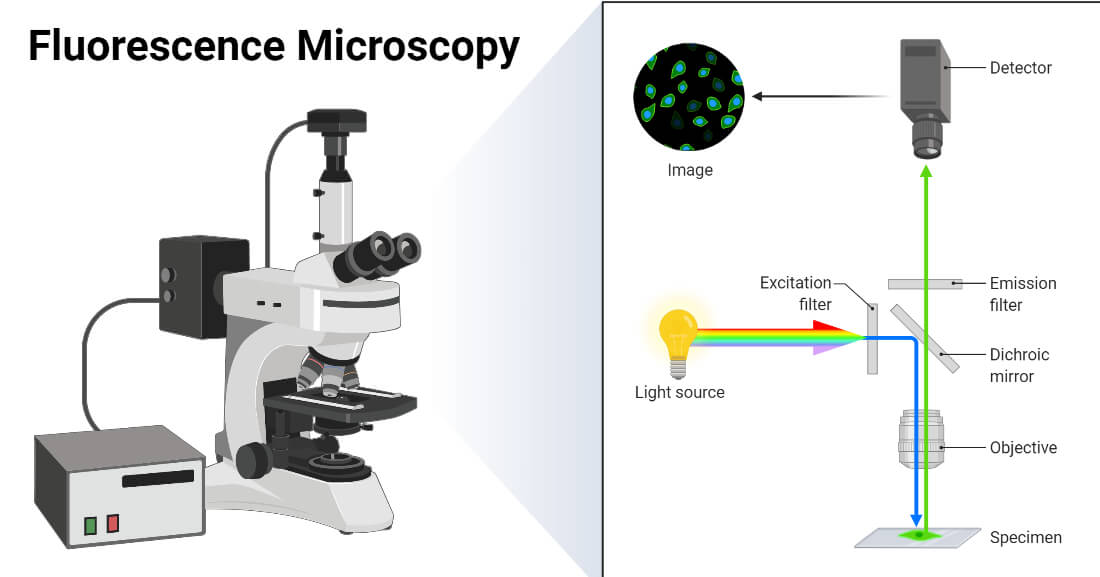
Fluorescent labels and light microscopy
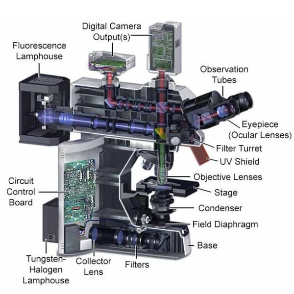
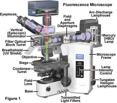
Fluorescence microscope - Wikipedia The majority of fluorescence microscopes, especially those used in the life sciences, are of the epifluorescence design shown in the diagram.Light of the excitation wavelength illuminates the specimen through the objective lens. The fluorescence emitted by the specimen is focused to the detector by the same objective that is used for the excitation which for greater resolution will need ... Fluorescence Imaging - Teledyne Photometrics Fluorescent molecules (known as fluorophores) are used to label samples, and fluorophores are available that emit light in virtually any color. In a fluorescent microscope, a sample is labeled with a fluorophore, and then a bright light ( excitation light) is used to illuminate the sample, which gives off fluorescence ( emission light ). How do you fluorescently label mRNA for microscopy? The vendor may be able to prepare fluorescently labeled mRNA for you; fluorescently-labeled nucleotides can simply be substituted during transcription. However, you can also do fluorescence in situ...
Fluorescent labels and light microscopy. Novel Fluorescent Label Shines a Light on DNA Structure in Cancer Cells Novel Fluorescent Label Shines a Light on DNA Structure in Cancer Cells, March 7, 2022, Researchers have developed a new fluorescent label that gives a clearer picture of how DNA architecture is... Fluorescence Microscope: Principle, Types, Applications Fluorescence microscopy is a light microscope that works on the principle of fluorescence. A substance is said to be fluorescent when it absorbs the energy of invisible shorter wavelength radiation (such as UV light) and emits longer wavelength radiation of visible light (such as green or red light). This phenomenon, also called fluorescence ... EYFP :: Fluorescent Protein Database Journal of Microscopy, 217(3) , 200-204. doi: 10.1111/j.1365-2818.2005.01437.x. Article Pubmed Partitioning of Lipid-Modified Monomeric GFPs into Membrane Microdomains of Live Cells Imaging Flies by Fluorescence Microscopy: Principles, Technologies, and ... Fluorescence microscopy in combination with specific labeling methods [ e.g., antibodies or fluorescent proteins (FPs)] enables selective visualization of the components of living matter, from molecules and organelles to cells and tissues, in both fixed and living organisms, and with high signal-to-noise ratio (SNR).
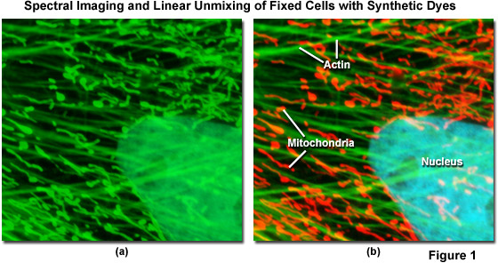
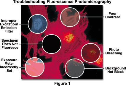
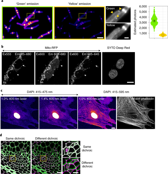
Light Sheet Fluorescence Microscopy Applications for Multicellular ... Light-sheet fluorescence microscopy has been shown to fit in this technological niche as it provides high optical resolution with 3D-imaging capabilities in large samples beyond several millimeters and up to several centimeters, while allowing the use of regular fluorescent labels or fusion proteins. Publications – Pradeep Research Group 2 days ago · Green Synthesis of Protein-Protected Fluorescent Gold Nanoclusters (AuNCs): Reducing the Size of AuNCs by Partially Occupying the Ca 2+ Site by La 3+ in Apo-α-Lactalbumin, Deepthi Yarramala, Ananya Baksi, Thalappil Pradeep and Chebrolu Rao, ACS Sustainable Chem. Eng., 5 (2017)6064-6069 (DOI: 10.1021/acssuschemeng.7b00958). Spectral Bleed-Through Artifacts in Confocal Microscopy - Olympus Imaging specimens having two or more fluorescent labels (or plant tissue sections exhibiting a high degree of autofluorescence) is often complicated by the bleed-through or crossover of fluorescence emission, unless the spectral profiles of the fluorophores are very well separated. ... Widefield microscopes equipped with LED light sources ... Fluorescent Labeling - What You Should Know - PromoCell Fluorescence microscopy allows the identification of cells and cellular components and the monitoring of cell physiology with high specificity. Fluorescence microscopy separates emitted light from excitation light using optical filters. The use of two indicators also allows the simultaneous observation of different biomolecules at the same time.
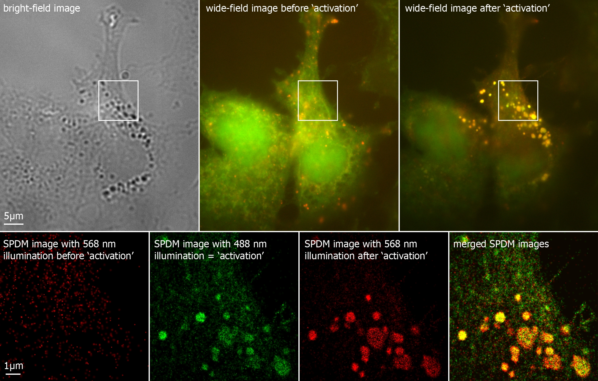
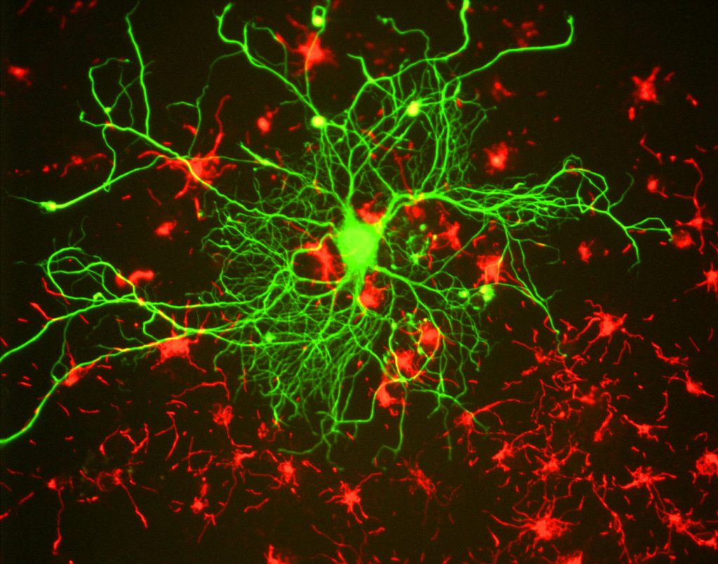
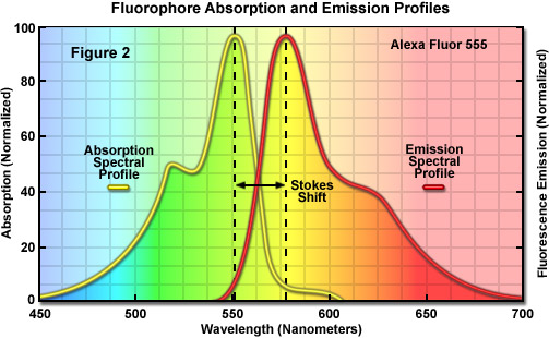
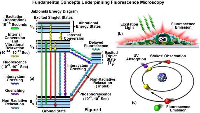
New fluorescent label provides a clearer picture of how DNA ... Unlike traditional fluorescence microscopy, which uses labels that glow constantly, this approach involves switching on only a subset of the labels at each moment. ... Discovery of 380-million ... Fluorescence Microscopy - Explanation and Labelled Images Fluorescence microscopy uses a high-intensity light source that excites a fluorescent molecule called a fluorophore in the sample observed. The samples are labeled with fluorophore where they absorb the high-intensity light from the source and emit a lower energy light of longer wavelength. Fluorescence Microscopy vs. Light Microscopy - News-Medical.net The fluorescent substances are called fluorophores, which are integrated into the sample. The fluorophores are excited by the light in the microscope, which causes them to give off light with lower... Fluorescence - Wikipedia Fluorescence is the emission of light by a substance that has absorbed light or other electromagnetic radiation.It is a form of luminescence.In most cases, the emitted light has a longer wavelength, and therefore a lower photon energy, than the absorbed radiation.
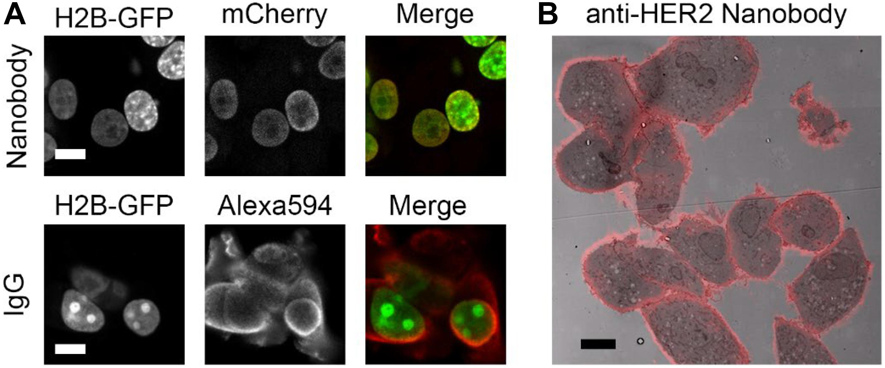
Immunolabeling for Correlative Light and Electron Microscopy on ... An ideal label for light and electron microscopy would contain both fluorescent dye and a colloidal nanoparticle on a single antibody molecule. Secondary and tertiary label, the colloidal nanoparticles are placed on the primary antibody while the fluorescent dye conjugated to secondary (D) or tertiary (E) antibody.
Fluorescent labeling of abundant reactive entities (FLARE) for cleared ... Fluorescence microscopy is a vital tool in biomedical research but faces considerable challenges in achieving uniform or bright labeling. For instance, fluorescent proteins are limited to model...
Important biomedical microscopy technique can now image ... - ScienceDaily The light needed to image fluorescent labels can be damaging and even deadly to delicate biological samples such as brain tissue or animal embryos used to study development and disease processes ...
In Silico Labeling: Predicting Fluorescent Labels in Unlabeled Images Microscopy is a central method in life sciences. Many popular methods, such as antibody labeling, are used to add physical fluorescent labels to specific cellular constituents. ... (ISL), reliably predicts some fluorescent labels from transmitted-light images of unlabeled fixed or live biological samples. ISL predicts a range of labels, such as ...
Dots, Probes and Proteins: Fluorescent Labels for Microscopy and Imaging Covering the spectrum, In essence, fluorescent probes are usually conjugated antibodies which are labelled (or 'tagged') with fluorophores. The two most common fluorophores used were FITC (fluorescein isothiocyanate) and TRITC (tetramethyl rhodamine isothiocyanate).
Labeling the ER for Light and Fluorescence Microscopy Most of them are not 100% specific for the ER membrane and may label other organelles at varying concentrations and incubation times. ... C., Wang, P., Kriechbaumer, V. (2018). Labeling the ER for Light and Fluorescence Microscopy. In: Hawes, C., Kriechbaumer, V. (eds) The Plant Endoplasmic Reticulum . Methods in Molecular Biology, vol 1691 ...
Fluorescent tag - Wikipedia Fluorescent tag. S. cerevisiae septins revealed with fluorescent microscopy utilizing fluorescent labeling. In molecular biology and biotechnology, a fluorescent tag, also known as a fluorescent label or fluorescent probe, is a molecule that is attached chemically to aid in the detection of a biomolecule such as a protein, antibody, or amino acid.
mNeonGreen :: Fluorescent Protein Database Sep 30, 2021 · mNeonGreen is the brightest monomeric green or yellow fluorescent protein yet described to our knowledge, performs exceptionally well as a fusion tag for traditional imaging as well as stochastic single-molecule superresolution imaging and is an excellent fluorescence resonance energy transfer (FRET) acceptor for the newest cyan fluorescent proteins.
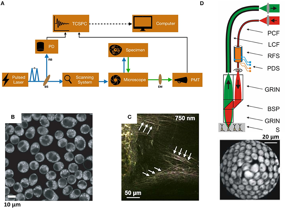
Fluorescence Microscopy & Cell Imaging | Research | UNM Cancer Center Fluorescence microscopy is routinely used to determine spatial and topological information about cells and tissues. Sophisticated laser scanning microscopic instrumentation, ultra sensitive digital cameras and specialized fluorescence probes make it possible to visualize cellular events in real time down to the molecular level.
Fluorescent Microscopy The basic task of the fluorescence microscope is to let excitation light radiate the specimen and then sort out the much weaker emitted light from the image. First, the microscope has a filter that only lets through radiation with the specific wavelength that matches your fluorescing material.
Semiconductor Nanocrystals as Fluorescent Biological Labels Sep 25, 1998 · In addition, nanocrystal probes may prove useful for other contrast mechanisms such as x-ray fluorescence, x-ray absorption, electron microscopy, and scintillation proximity imaging, and the use of far red– or infrared-emitting nanocrystals (InP and InAs) as tunable, robust infrared dyes is another possibility.
Researchers demonstrate label-free super-resolution microscopy - Phys.org A newly developed sub-diffraction-limit microscopy approach doesn't require fluorescent labels. The video shows the process of the data evaluation algorithm, retrieving the positions and sizes of...
Introduction to Fluorescent Proteins | Nikon’s MicroscopyU The brightness and fluorescence emission spectrum of enhanced yellow fluorescent protein combine to make this probe an excellent candidate for multicolor imaging experiments in fluorescence microscopy. Enhanced yellow fluorescent protein is also useful for energy transfer experiments when paired with enhanced cyan fluorescent protein (ECFP) or ...
Labeling Proteins For Single Molecule Imaging - Teledyne Photometrics Most of the fluorescent dyes used in microscopy have certain wanted and unwanted characteristics. Usually, they get excited by a lower wavelength (higher energy light), get elevated from a ground state to an excited state and - while falling back to the ground state - emit photons of a longer wavelength (lower energy light).
Department Hell | Max Planck Institute for Multidisciplinary ... Dec 03, 2021 · To surpass the diffraction barrier, all these methods utilize a reversible transition or switch of fluorescent labels between a bright and a dark state. In combination with 4Pi microscopy, which is another concept developed by this group that uses two opposing lenses, the resolution can be increased in all spatial dimensions down to the ...
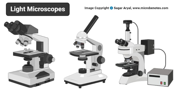
Light Microscope- Definition, Principle, Types, Parts, Labeled Diagram ... A light microscope is a biology laboratory instrument or tool, that uses visible light to detect and magnify very small objects and enlarge them. They use lenses to focus light on the specimen, magnifying it thus producing an image. The specimen is normally placed close to the microscopic lens.
Label-Free Multiphoton Microscopy: Much More Than Fancy Images Light microscopy is a gold standard technique in biomedical research and clinical diagnosis . ... Sapphire laser with an OPO allows multiplexed fluorescence and label-free analysis of the same sample, as shown in Figure 2 d on a lung ex vivo sample. 3. Label-Free Multiphoton Microscopy in Biomedical Research
Multispectral intravital microscopy for simultaneous bright-field and ... Conventional light microscopes do not allow for simultaneous bright-field and fluorescent imaging. Moreover, in conventional microscopes, only one type of fluorescent label can be observed. This study introduces multispectral intravital video microscopy, which combines bright-field and fluorescence microscopy in a standard light microscope.
How do you fluorescently label mRNA for microscopy? The vendor may be able to prepare fluorescently labeled mRNA for you; fluorescently-labeled nucleotides can simply be substituted during transcription. However, you can also do fluorescence in situ...
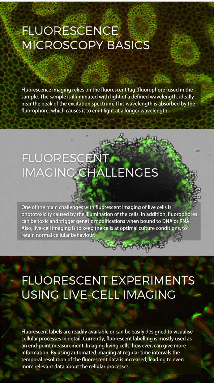
Fluorescence Imaging - Teledyne Photometrics Fluorescent molecules (known as fluorophores) are used to label samples, and fluorophores are available that emit light in virtually any color. In a fluorescent microscope, a sample is labeled with a fluorophore, and then a bright light ( excitation light) is used to illuminate the sample, which gives off fluorescence ( emission light ).
Fluorescence microscope - Wikipedia The majority of fluorescence microscopes, especially those used in the life sciences, are of the epifluorescence design shown in the diagram.Light of the excitation wavelength illuminates the specimen through the objective lens. The fluorescence emitted by the specimen is focused to the detector by the same objective that is used for the excitation which for greater resolution will need ...

















Post a Comment for "43 fluorescent labels and light microscopy"